Knee
Knee Pain and Conditions
The knee is the strongest joint in the human body, allowing the legs to bend and straighten while carrying almost all of the weight of the individual when they are standing. The knees are a hinge joint but still have substantial capacity for lateral (side-to-side) motion.
As an active, weight-bearing joint, the knee is a source of pain and problems for many people. This pain may be acute or chronic, and may be a result of injury, overuse, or growth. It can stem from the tendons, ligaments, bones, cartilage, or any other structure within the knee.
When surgery is indicated, Orthopaedics New England specializes in the most advanced, cutting-edge procedures to help our patients get back to their desired level of activity as quickly as possible. Learn more about the knee below and treatments we provide for knee injuries and diseases.
To schedule an appointment with one of our orthopedic knee surgeons, call (203) 598-0700 or use our online form. We have three offices to serve you in Middlebury, New Milford, and Farmington, Connecticut.
Knee Anatomy
Knee Diseases
Knee Arthritis
Non-Operative Knee Treatment
Knee Replacement
Knee Anatomy
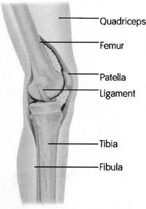 The knee joins together the lower end of the femur (thigh bone) and the upper end of the tibia (the large bone in the lower leg; the smaller one is the fibula). The patella (knee cap) also is in contact with the femur, where it glides up and down in a shallow trough called the trochlea.
The ends of the bones and the undersurface of the patella are covered with a layer of articular cartilage that is similar to the Teflon coating in a frying pan. The coating is normally about 1/8th of an inch thick, but over time, wear and tear and a number of diseases, such as osteoarthritis, can lead to the loss of this cartilage coating. When the cartilage is worn away, most patients experience significant and worsening pain from the bone-on-bone contact.
The knee joint is surrounded by a capsule. There is a tough membrane called the retinaculum that covers it along the front, going off either side of the patella. Sometimes this retinaculum can develop tears in an injury, and the patella may not track correctly afterwards as a result.
The knee joint has three compartments within it. Two of these are formed where the curved, cam-shaped end of the femur meets the tibia on both the inner (medial) and outer (lateral) side of the knee. The third compartment is formed by the patella and the trochlear groove of the femur that it glides up and down in.
Knee problems can occur in any and all of these three compartments. Meniscal problems happen in the medial and lateral compartments, where most of the weight bearing also occurs. Problems with the undersurface of the patella (e.g., chondromalacia patellae) manifest in the patellofemoral compartment.
Sometimes arthritis and other conditions affect only one side of the knee, or affect it to a much greater degree than the rest of the knee, and therefore partial knee replacements and other procedures may focus on just that compartment.
At other times, the term tricompartmental degenerative joint disease is used to describe arthritic changes affecting all three compartments, which may dictate a total knee replacement instead of a partial one.
Ligaments are strong tissues that provide stability and allow motion. They enable your knees to have the flexibility to move in various directions while maintaining balance. There are four primary ligaments around the knee that hold it together:
- The anterior cruciate ligament (ACL) and posterior cruciate ligament (PCL) cross inside of the knee, connecting the femur to the tibia. Together they counteract excessive forward and backward forces and prohibit displacement of the bones. The ACL is the most commonly injured ligament in the knee.
- Located on the inner (medial) and outer (lateral) sides of the knee, the medial collateral ligament (MCL) and lateral collateral ligament (LCL) help the knee joint resist side-to-side stress and maintain position.
Within the space between the femur and tibia are two cartilage structures called menisci (the singular term is meniscus), one in each side of the knee (medial and lateral). These C-shaped gaskets are made of the same type of cartilage as your nose and ears. They fit around the rim of the knee and function as shock absorbers.
Years ago, painful meniscus tears were typically treated by removing the entire meniscus. However, this eventually led to significant arthritis. Today these tears are treated with arthroscopic surgery.
The quadriceps, the largest muscle group in the thigh, attaches to the end of the patella (kneecap). Below the patella is a tough tendon that connects the patella to the front of the tibia. Together these structures function like a pulley, and when the thigh muscles contract, the tibia is extended and the knee straightens.
Any injury to these structures can be devastating. Quadriceps tears, patella fractures, and patella tendon ruptures are all usually repaired surgically because of the importance of this entire assembly in moving the knee backwards and forwards.
Knee Diseases
Osteoarthritis is the most common cause of knee pain. Other causes of knee pain include meniscal tears, knee bursitis/tendinitis, ligament injuries, osteochondral defects/loose bodies, and chondromalacia.
The tendons around the knee can develop inflammation as a result of overuse, injury, structural abnormalities, or diseases such as arthritis. While tendinitis usually resolves with rest and physical therapy, in severe cases the quadriceps or patella tendon can rupture, requiring surgical repair.
There are several bursae around the knee joint. These fluid-filled sacs help muscle layers slide smoothly over each other, but the sac can sometimes become inflamed and painful. Overuse, trauma, chronic arthritis, degenerative joint disease, and obesity, especially in middle-aged women, are frequent causes of knee bursitis. It is typically treated with steroid injections, anti-inflammatory medication, and physical therapy.
Sometimes a plug of cartilage and underlying bone can break loose within the knee joint, leaving a crater in the normally smooth surface. This crater by itself is harmful and often leads to accelerated arthritis, but the chunk of bone and cartilage (osteochondral) can be even more painful and destructive as it moves around inside the knee joint. This loose body can cause the knee to catch and lock. Arthroscopic surgery is usually required to remove the defect.
When articular cartilage begins to delaminate or peel away from the bone, this process is known as chondromalacia. It doesn’t mean that serious arthritis is imminent; it just indicates that degenerative changes are probable down the road. Worn cartilage can be shaved away during arthroscopic surgery.